Showing posts with label disease. Show all posts
Wednesday, November 2, 2016
CHF
CHF is caused by fluid overload in the heart and. CHF is totally different from COPD. COPD is all about the lungs. Why is it called congestive heart failure? Because it's so much fluid. What kills HF pt? How does it kill the pt? It's the hypoxia. The real immediate problem is hypoxia, not fluid overload. It's not how much fluid int the body but it's how much fluid in the lungs. When the pt is having HF, that's the main problem. Why is there fluid in the lungs? Congestion means it's referring to the lungs. If you do a CXRAY, you'll see big heart. What's congestion? What do you see on cxray? You'll see big heart but that's not important, what's important? FLUID IN THE LUNGS. You'll see pulmonary edema = fluid overload/cushiness/leaking out in the lungs. Pulmonary edema = that's what you'll see. When CHF pt comes in, the real problem is hypoxia. You can help solve that and makes that better by giving oxygen. But ultimately, you also have to know the root cause. And, the root cause or the cause is pulmonary edema. PULMONARY EDEMA. So, what we need to know is then to get the fluid out of hte lungs. When the pt is SOB and hypoxic, do you care about getting the fluid out of the legs? NO, you care about getting the fluids out of the lungs: you want to get the pt's legs down ont he ground. Is it going to get their fluid in the legs worse? Yes. What's the problem? The LEFT ventricle. The left ventricle takes blood from the lungs and sends to the rest of the body. The right ventricle tkes blood from the rest of the body and sends it to the lungs. So, CHF, we have blood going to the lungs but we can't pump it out. There's fluid in lungs. Our main problem is the Left ventricle is not strong enough to pump fluid out. So if pt has CHF, FLUID IN THE LUNGS = HYPOXIC. And, if I make the Rt ventricle stronger, will that make the problem better or worse? worse. They may have some weakness in the Rt ventricle but the main problem is the Left ventricle is not effective enuf to pump the blood out. So, the way we can treat that is that we can make the heart stronger. This is a temporary fix. Slowing the gas pedal when the engine is already not doing so well. So, the meds like digoxin and dopamine and dobutamine will stimulate the heart and cause the heart to pump more out. If the heart is okay, it will regulate itself and it will try to push the left side harder to get that fluid out. So, how do we address it otherwise? We give them meds that cause pulling blood in the legs? Stand them up, put the legs down, use gravity. You can give the NTG. What does NTG do? It's a vasodilator. If I vasodilate my peripheral vein, what does that do? I pull more fluid out. It will only go so far tho. Bc you didn't get rid of any fluid in the body but you just move it around to somewhere else. How do I get the fluid out of the body? You actually have to use diuretics. If there's less fluid in the body, there's more fluid going to the Rt ventricle or what? Less. The Rt ventricle is going to pump less. And, the Lt ventricle job easier or harder? Easier.
What's the most common cause of CHF? HTN is a common cause. Sure. What else? too much IV fluid - might be a cause acutely in a pt in a hospital and unable to control their fluid bc their doctor/nurse is giving too much fluid but usuaully not in the world. Why does the pt have a weak heart? Not smoking but smoking makes it worse. Kidney failure? pretty close. CORONARY ARTERY DISEASE due to recurrent MI and hyperlipidemia. The 2 most common causes of CHF = CORONARY ARTERY DISEASE due to recurrent ischemia to the heart and infarction which kill the vessels in the heart and make the heart thin and dilated too large and weak. The other common cause of CHF in older ppl but not until they're very old is HTN leading to thickening of the heart bc the heart to work hard to push the BP so it gets bigger and thicker and the ventricle cavity smaller. And, they have diastolic or systolic HF? DIASTOLIC. Why diastolic HF? Why? bc it's too thick and it cannot dilate. It's old heart and it's thick. Main issue is poor diastolic function. This is a particular common cause in pt who's older and long-standing HTN. So, if 88 y/o women comes to clinic with HF and she never smokes and she hasn't had many medical problems all her life. She's probably has diastolic HF. If 48 y/o african american man, who eats meat smokes and drinks gambles and does drugs doesn't take care of himself and comes to ur clinic and hospitalized, coronary artery disaese HF, primarily systolic HF bc the heart is too thin so it cannot pump.
PE: edema in legs. Hears fluid in the lungs (crackles).
~bf
COPD
COPD usually caused by smoking. COPD = chronic obstructive pulmonary disease. Chronic means >6mo. Obstructive means there's a blockage. Pulmonary means the lungs. So, COPD is the obstructive dz of the lungs. We used to call it emphysema = damage of the alveoli, they are very thin. They don't have much air exchange. They kinda thin out and they get bigger. Lungs is like sponge = tiny little holes and pockets for air to move around. It looks like sponge. It should look like a sponge. COPD lungs are mostly air. And, that's bad. We don't want lungs to be full of air. The lungs should be a sponge. COPD lungs and chest x-ray, it's much more overinflated and clear. U should be able to see it on CXRAY. It's going to look more black.
They also used to be chronic bronchitis. Chronic inflammation of the bronchi. They get narrowed and they get full of junk and they get narrower and they get infxn. They can have some phlegm and coughing and can be like asthma.
When the pt is having a very difficulty time breathing and they try to exhale, it doesn't work. And, that doesn't work. You'll see pt trying to breathe out slowly. They have prolonged expiratory phase. They breathe out very long and slowly with their mouth closed. That's what they call purse lip breathing. They do this to compensate for obstructive bronchi.
Now we know that it's not one thing or another. Most pt have emphysema or chronic bronchitis. Most ppl have features of both. So, hyper-inflated lungs, difficulty breathing, coughing, more infxn, chronic. Do their lungs get oxygen better or worse? Worse? Because damage of alveoli and obstructive bronchi. What's their pulse ox going to be ? Less than normal.
PE: no fluid sounds in lungs (no crackles). but hyper-resonating sounds (egophony).
They also used to be chronic bronchitis. Chronic inflammation of the bronchi. They get narrowed and they get full of junk and they get narrower and they get infxn. They can have some phlegm and coughing and can be like asthma.
When the pt is having a very difficulty time breathing and they try to exhale, it doesn't work. And, that doesn't work. You'll see pt trying to breathe out slowly. They have prolonged expiratory phase. They breathe out very long and slowly with their mouth closed. That's what they call purse lip breathing. They do this to compensate for obstructive bronchi.
Now we know that it's not one thing or another. Most pt have emphysema or chronic bronchitis. Most ppl have features of both. So, hyper-inflated lungs, difficulty breathing, coughing, more infxn, chronic. Do their lungs get oxygen better or worse? Worse? Because damage of alveoli and obstructive bronchi. What's their pulse ox going to be ? Less than normal.
PE: no fluid sounds in lungs (no crackles). but hyper-resonating sounds (egophony).
atelectasis
it can be normal or it can be severe or bad. A lot of time some atelectasis is normal if you've been in bed all day and not getting around. That's why you need the pt to take a deep breath a couple times before you do the pulse ox. If they've been in bed all day, have them take a couple of breaths to clear out atelectasis. Atelectasis has nothing to do smoking. It's usually due to pt staying in bed all day.
Wednesday, September 14, 2016
Barrett's esophagus surveillance
- No dysplasia - 2 EGD w/ biopsy within 1 year
+If negative, f/u EGD in 3 yrs
- Low-grade dysplasia*
+Biopsy in 6 months
+Repeat EGD qyear until no dysplasia x 2
- High-grade dysplasia*
+Repeat EGD with biopsy q3months + send to ER
+Surveillance q3months with EGD
*Dx dysplasia should be confirmed with 1 additional pathologist, preferably an expert in esophageal histopathology
+If negative, f/u EGD in 3 yrs
- Low-grade dysplasia*
+Biopsy in 6 months
+Repeat EGD qyear until no dysplasia x 2
- High-grade dysplasia*
+Repeat EGD with biopsy q3months + send to ER
+Surveillance q3months with EGD
*Dx dysplasia should be confirmed with 1 additional pathologist, preferably an expert in esophageal histopathology
Barrett's esophagus screening
Screening for BE may be considered in men with chronic (>5 years) and/or frequent (weekly or more) symptoms of gastroesophageal reflux (heartburn or acid regurgitation) and two or more risk factors for BE or EAC. These risk factors include: age >50 years, Caucasian race, presence of central obesity (waist circumference >102 cm or waist–hip ratio >0.9), current or past history of smoking, and a confirmed family history of BE or EAC (in a first-degree relative) (strong recommendation, moderate level of evidence).
Given the substantially lower risk of EAC in females with chronic GER symptoms (when compared with males), screening for BE in females is not recommended. However, screening could be considered in individual cases as determined by the presence of multiple risk factors for BE or EAC (age >50 years, Caucasian race, chronic and/or frequent GERD, central obesity: waist circumference >88 cm, waist–hip ratio >0.8, current or past history of smoking, and a confi rmed family history of BE or EAC (in a fi rst-degree relative)). (strong recommendation, low level of evidence).
Screening of the general population is not recommended (conditional recommendation, low level of evidence).
Before screening is performed, the overall life expectancy of the patient should be considered, and subsequent
See more:
http://gi.org/wp-content/uploads/2015/11/ACG-2015-Barretts-Esophagus-Guideline.pdf
http://www.medscape.com/viewarticle/854725
Given the substantially lower risk of EAC in females with chronic GER symptoms (when compared with males), screening for BE in females is not recommended. However, screening could be considered in individual cases as determined by the presence of multiple risk factors for BE or EAC (age >50 years, Caucasian race, chronic and/or frequent GERD, central obesity: waist circumference >88 cm, waist–hip ratio >0.8, current or past history of smoking, and a confi rmed family history of BE or EAC (in a fi rst-degree relative)). (strong recommendation, low level of evidence).
Screening of the general population is not recommended (conditional recommendation, low level of evidence).
Before screening is performed, the overall life expectancy of the patient should be considered, and subsequent
See more:
http://gi.org/wp-content/uploads/2015/11/ACG-2015-Barretts-Esophagus-Guideline.pdf
http://www.medscape.com/viewarticle/854725
Barrett's esophagus
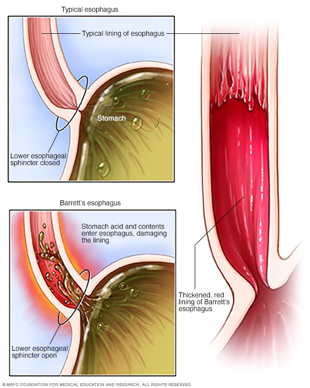
In Barrett's esophagus, normal esophageal cells are replaced with abnormal cells. Barrett's esophagus is thought to be caused by long-standing gastroesophageal reflux disease (GERD), which causes stomach contents to back up into the esophagus.
In Barrett's esophagus, tissue in the tube connecting your mouth and stomach (esophagus) is replaced by tissue similar to the intestinal lining.
Barrett's esophagus is most often diagnosed in people who have long-term gastroesophageal reflux disease (GERD) — a chronic regurgitation of acid from the stomach into the lower esophagus. Only a small percentage of people with GERD will develop Barrett's esophagus.
Barrett's esophagus is associated with an increased risk of developing esophageal cancer. Although the risk is small, it's important to have regular checkups for precancerous cells. If precancerous cells are discovered, they can be treated to prevent esophageal cancer.
Chronic constipation
Dx test: Anorectal manometry to r/o Hirschsprung's & to aid dx of fecal incontinence.
Saturday, April 2, 2016
acromegaly due to pituitary tumor
**All cases are fictitious. No real patient encounters. Any resemblance to real persons, living or dead, is purely coincidental.**
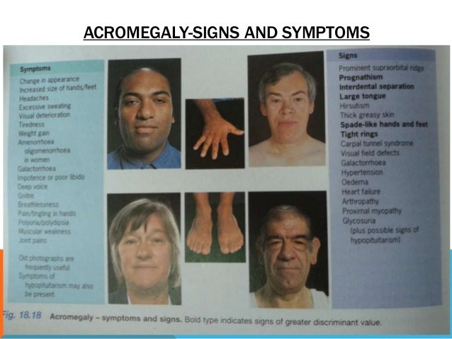
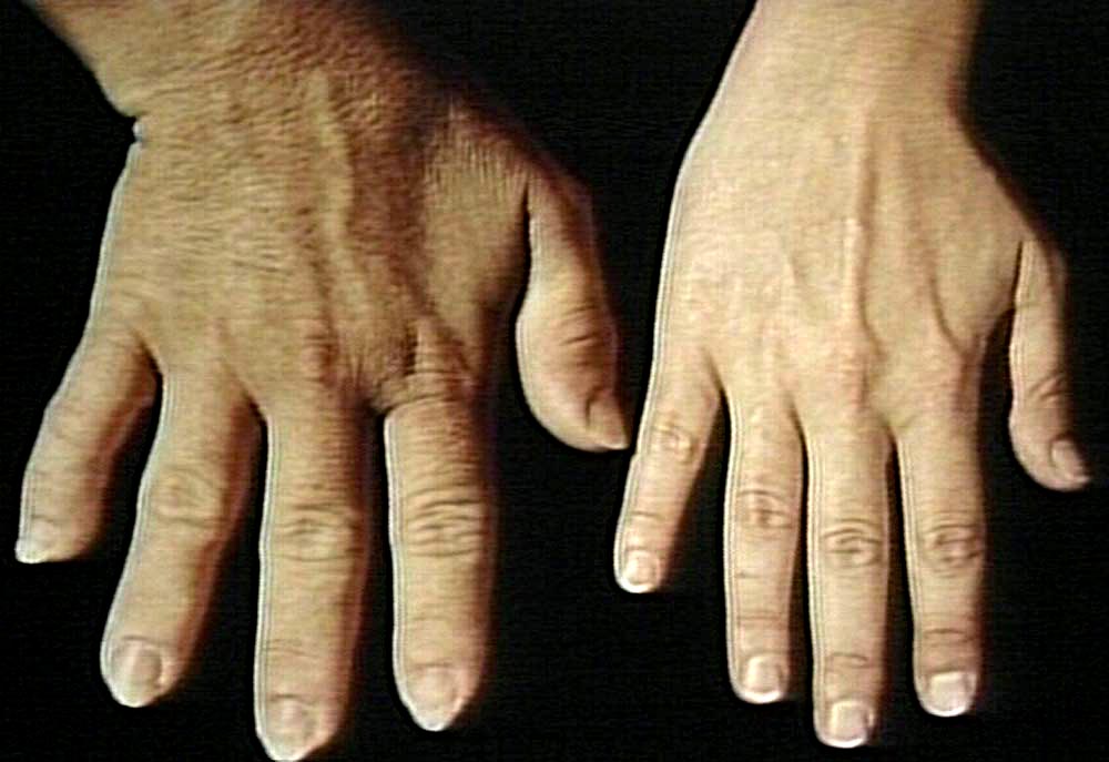
Case*: Oh my, his eye brow bone is huge!
Thinking: What could be the cause?
Case*: Oh my, his eye brow bone is huge!
Thinking: What could be the cause?
parkinsonism
Facial masking: blank expressions, immobilized, staring
Shuffling gait: walk by dragging one's feet along or without lifting them fully from the ground. Turning is en bloc like a statue.
Pill-rolling tremors: "the motion used to roll a marble between the thumb and forefinger"
"The tremor often spreads to the other side of the body as the disease progresses, but will remain most obvious on the side of the body where it first started.A few more points on tremors include:
- usually occur at rest, may occur at any time
- may become severe enough to interfere with activities
- may be worse when tired, excited, or stressed
- finger-thumb rubbing (*pill-rolling tremor) may be present
*NOTE: ("Pill-rolling" is seen especially in the hands; this is fairly unique to Parkinson's disease. The term refers to the motion that a pharmacist uses to align a handful of pills before placing them in a bottle or, possibly, the motion used to roll a marble between the thumb and forefinger. Eventually the tremor becomes more generalized.)"
Thursday, March 31, 2016
Hepatitis A
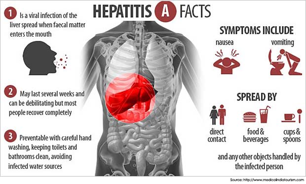
Mini-Case*: Pt's Hepatitis A came back positive results. Why don't you treat patient for Hepatitis A?
Thinking: it's positive. Why don't you treat?
Conclusion: Self-limited (look up self-limited in medical dictionary if you don't know what it is already). Supportive therapy only. Excellent prognosis.
Resources:
Helpful chart to intepret different types of hepatitis lab test results: http://oaml.com/PDF/2010/Hepatitis%20Results%20Interp.%20FINAL-Aug%2024%202010.pdf
Thinking: it's positive. Why don't you treat?
Conclusion: Self-limited (look up self-limited in medical dictionary if you don't know what it is already). Supportive therapy only. Excellent prognosis.
More:
When to order:
- To help dx acute hepatitis cause
- Evaluate the need for hep A vaccine
What to order:
- Part of viral hepatitis panel.
What to collect:
- Blood sample
Resources:
Helpful chart to intepret different types of hepatitis lab test results: http://oaml.com/PDF/2010/Hepatitis%20Results%20Interp.%20FINAL-Aug%2024%202010.pdf
Subscribe to:
Posts (Atom)