Showing posts with label ekg. Show all posts

Monday, December 26, 2016

EKG height


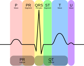
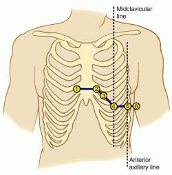
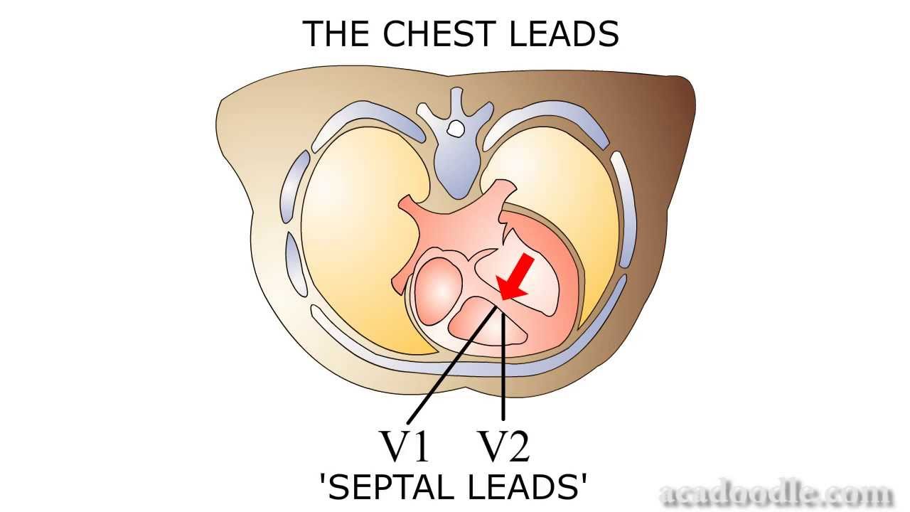
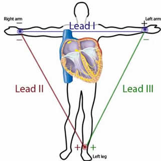
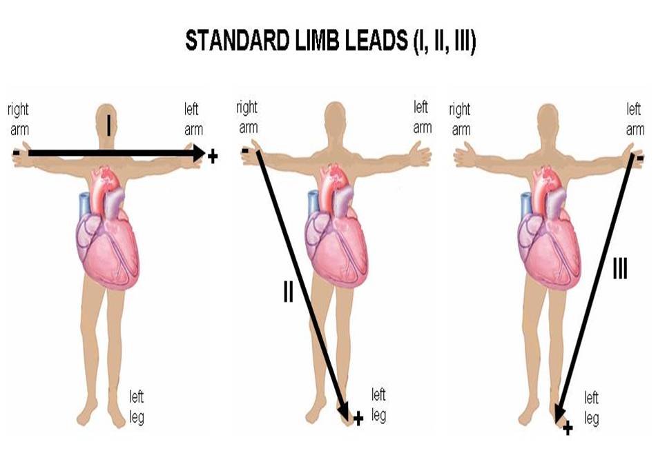
P waves in leads II and V1.
  •      If the P in lead II is greater than 2.5 mV (small boxes) right atrial enlargement (RAE) probably exists. 
  •       If the P wave in V1 is negative or biphasic, then LAE probably exists.  
    • This negative portion of the P wave in V1 should be more than 1 box wide and 1 box down to be considered significant.
PR segment
  • PR depression. The only classic significant PR segment abnormality that is encountered in the emergency department is PR depression.  This is most often seen in the setting of pericarditis.  Since there are multiple stages of pericarditis, these depressions are not always seen when this disease is present.
QRS
  • QRS width:
    • QRS >3ss (0.12 sec) = BBB 


  • QRS shape:
    • RBBB --- RSR' = rabbit ears = The “R” from Right and Rabbit = right BBB (RBBB) there should be an RSR’ in leads V1, V2 or V3.
    • LBBB --- a left BBB (LBBB) you need a deep, wide Q/S wave in these anterior leads.  This you will just have to memorize.


    •  QRS height:
      • LVH --- left ventricular hypertrophy (LVH)
        • A positive deflection in leads I or aVL greater than 11 mV. Mnemonics: 1 and L look like an 11 when they are side by side.
      

        • A value greater than 35 mV when you add the absolute values of the more negative of V1 or V2 plus the more positive of V5 or V6. Mnemonics: just have to look at the Q or S in leads V1 and V2 and see which is more negative.  Take the absolute size of that complex and add it to the larger R of V5 or V6.
        • Remember, only one criterion is sufficient to diagnose LVH.

      • ST segments:
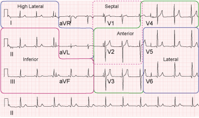
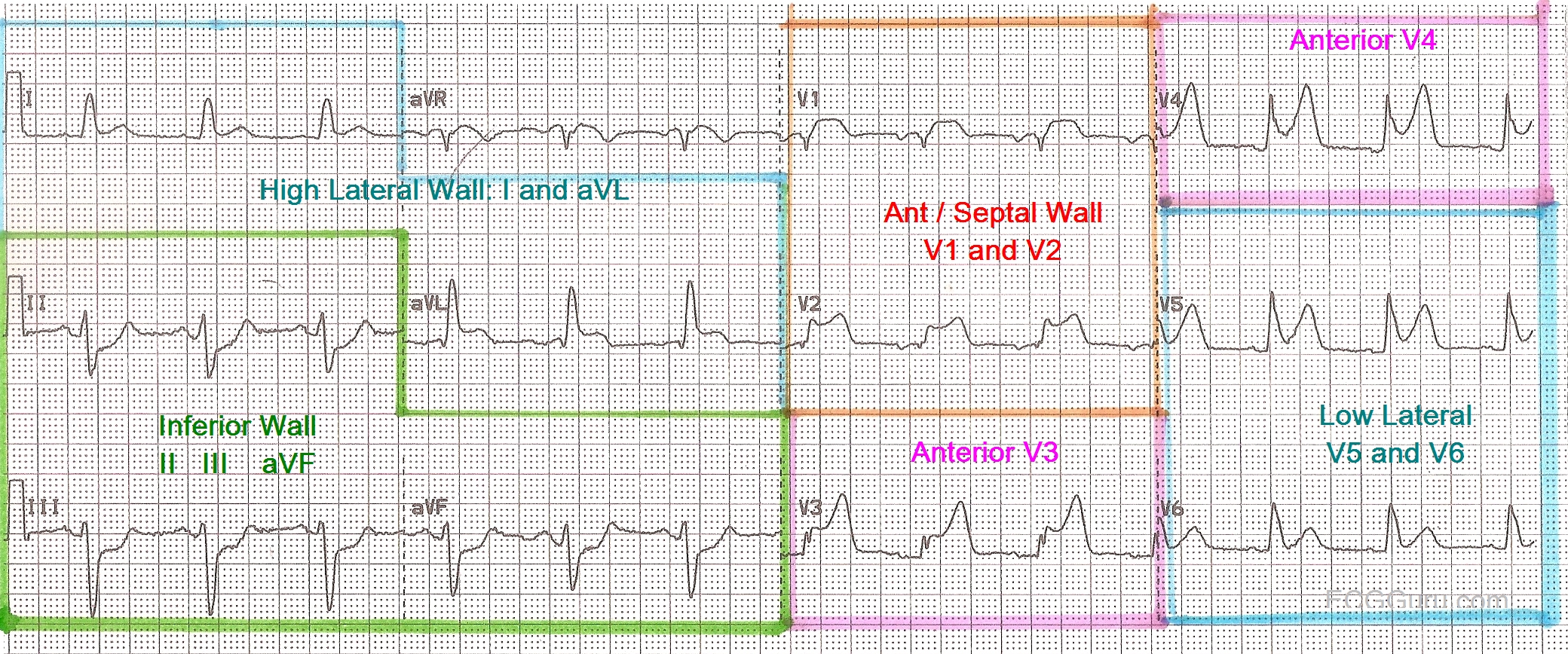
        • you need to check systematically through all 12 leads of the EKG looking for ST elevations or depressions.  These findings are consistent with AMI or ischemia respectively.


      • T wave:
        • Specifically, you are looking for flipped T waves that are pointing in the negative direction.  This is also symbolic of coronary ischemia.  
       
        • Quickly glance at the shape of the T waves.  If they are sharp and pointy instead of nicely rounded, hyperkalemia may exist. 

          EKG width

          P wave width = 0.12 sec (3ss) (>0.12 or >3ss <=> atrial enlargement)
          PR interval (from the beginning of P wave to 1st deflection of QRS) = or <  0.20 sec (5ss = 1ls)  (if >0.20 or 5ss <=> 1st degree AV block)


          QRS width = 0.12 sec (3ss) (>0.12 or 3ss <=> bundle branch block (BBB)  (partial or full) OR the complex did not take the conduction pathway at all and had a ventricular origin as is seen in a premature ventricular complex or PVC)

          QTc width < 450sec => If >450msec, it may be a tip-off that electrolyte abnormalities exist or some toxin is present.  While this concept is more complex than depicted here, it is not so important and can be discussed later when looking at specific EKG’s


          ekg basics


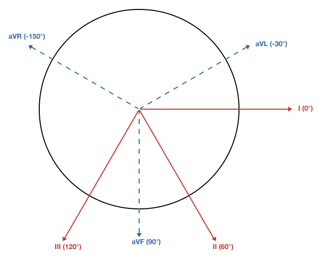
          ekg axis

          LAD

          ekg rhythm

          P wave - precedes every QRS?
          P wave morphology - same shape? - originates in every SA node
          PR interval - same length?


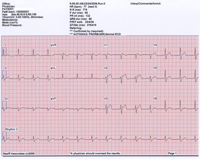
          Normal EKG


          ekg rate

          Mnemonics 300-150-100-75-60-50
          300/n


          300/1 = 300
          300/2 = 150
          300/3 = 100
          300/4 = 75
          300/5 = 60
          300/6 = 50


          Monday, September 26, 2016

          Atropine

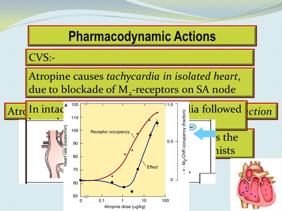
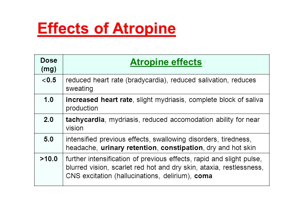
          - Atropine is a parasympatholytics that blocks the M2 receptor of the SA node. Therefore, atropine increases the rate of SA node firing & thus => improving the conduction thru the AV node.
          => Therefore, Atropine can be used for sinus bradycardia because the SA node just needs some kicks.
          => Therefore, Atropine can be used for 2nd degree AV block Wenckebach bc the site of the cause of the AV node wenckeback is at the AV node.
          => Therefore, Atropine is not effective for 2nd degree AV block Mobitz type 2 because the site of the problem blockage is below the AV node.


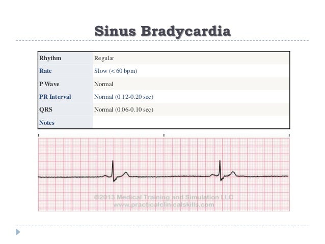
          sinus bradycardia


              Causes:
          - Electrolyte imbalances
          - Acid/Base problem
          - Hypoxia
          - Temporary or permanent pacing (LOOK FOR THEIR PACEMAKER. If they depend on their pacemaker to fire and it's not working, they probably have permanently slow heart rate! Need the pace maker to be fixed!)

          Tx:
          None if asymptompatic

          If symptomatic (hypotensive, keeps passing out), then:
          1 - atropine (atropine is a parasympatholytic, therefore, it will pace the heart making it beat faster; However, be careful not to use atropine on patients with MI because pt will go to V-Tach with the increased myocardial demand workload and oxygen,

          2 - pacer, (SA node is not working right, may need pacer)

          3 - r/o vagal problem esp. in children bc they like to hold their breath and thus getting into fainting issues a lot!)


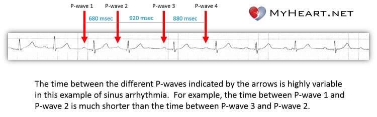
          sinus arrhythmias


          1 - Rate: fast and slow and slow and fast unpredictably

          2 - Rhythm:
          - P is before each QRS. QRS is after each P.
          - Q is normal looking
          - Irregular: there's like a pause between some beat

          3 - Axis: skip this for now

          4 - Width:
          - PR interval width is normal (5ss at all beats) and fixed (Remember sinus rhythm has fixed and normal PR interval)
          - Q width is normal (<3ss)
          - RR: 1/2RR width is less than QT segment= normal

          5 - Height:
          - No ST elevation
          - Upright P wave in lead II (Remember sinus rhythm has upright P wave in lead II)

          6 - Pts symptoms:
          - Usually EKG sinus arrythmias will have rhythms change as pt breathing in and breathing out
          - Usually older pt with syncope, fatigue and worsening HF may have sinus arrhythmias or sinus bradycardia NOT owing to use of beta blocker, that would mean pt may need a pacemaker (sick sinus syndrome)


          vfib

          Symptoms: pt not looking at u, totally out, no pulse, no breathing,
          EKG:

          Tx:
          CPR, defib, epinephrine, IV, oxygen, intubation, implantable defibrillator to prevent future incident

          Work up: Treatable causes: H's & T's

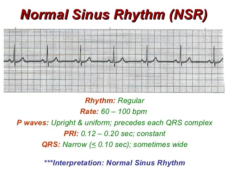
          NSR - Normal sinus rhythm

          Qt tip:  NSR ALWAYS has upright P wave with normal length PR.
          (assuming no dextrocardia or misplaced leads)


          Sunday, September 25, 2016