Showing posts with label anatomy. Show all posts

Saturday, April 2, 2016

ruptured tympanic membrane

**All cases are fictitious. No real patient encounters. Any resemblance to real persons, living or dead, is purely coincidental.**
 
Case*: hmm, I don't see the Tympanic membrane. Did I not push my otoscope far in enough?

Solution:

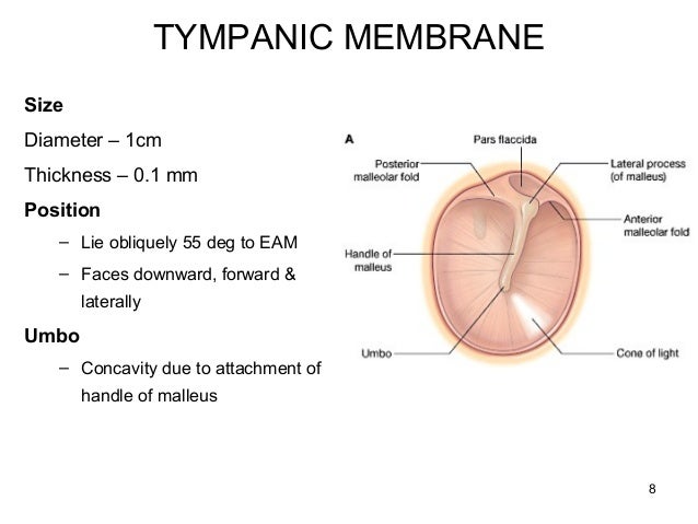
NORMAL TYMPANIC MEMBRANE:
Know what a normal tympanic membrane look like:
- pinkish gray (Wonder me! usually sees normal tympanic membrane more gray thank pinkish)
- small blood vessels along the malleus is normal too

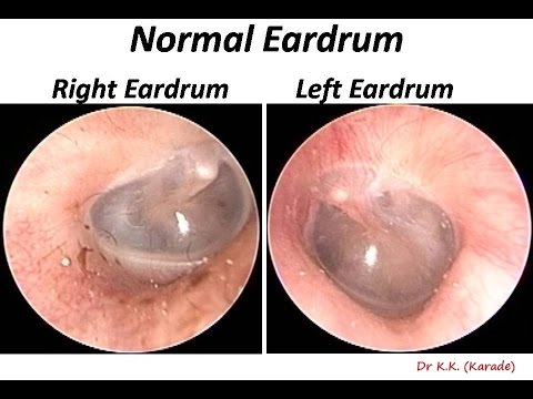

Note: malleus, pars flaccida, pars tensa, & cone of light

 
Malleus = Hammer


Ruptured tympanic membrane:

Helpful resource: http://www.ghorayeb.com/Malleus.html

Sunday, March 20, 2016

ankle anatomy


Ankle anatomy:
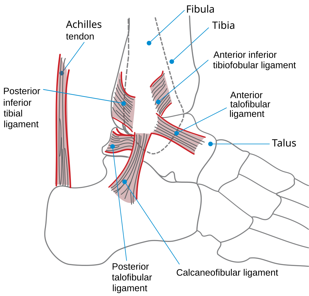
Lateral ankle ligaments: anterior talofibular ligament, posterior talofibular ligament, calcaneofibular ligament
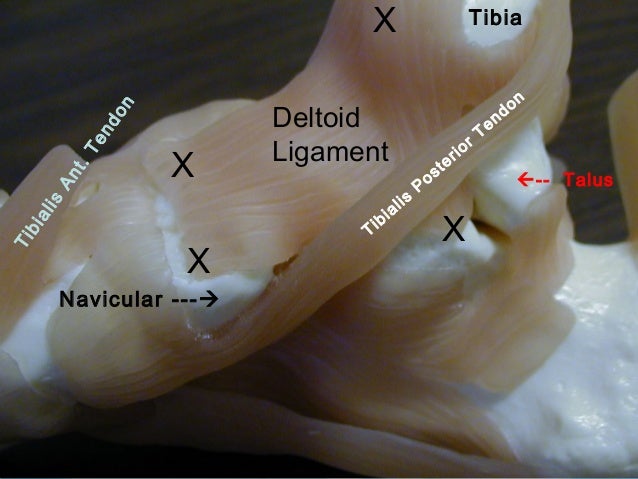
Medial ankle ligament: deltoid ligament
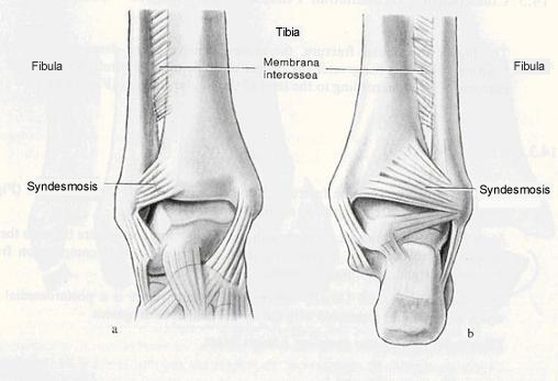
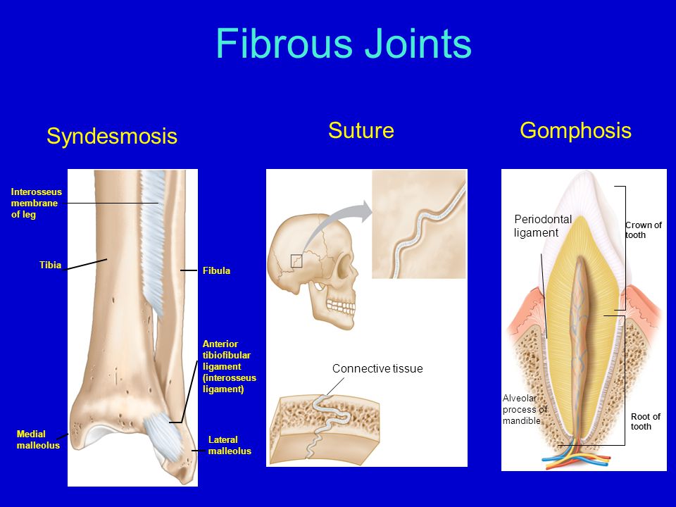
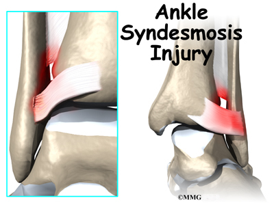
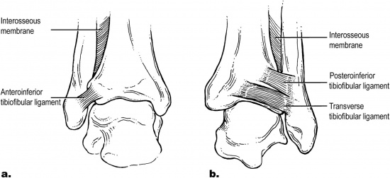
Tibiofibular ligaments/syndesmosis: anterior tibiofibular ligament, posterior tibiofibular ligament, interosseous membrane
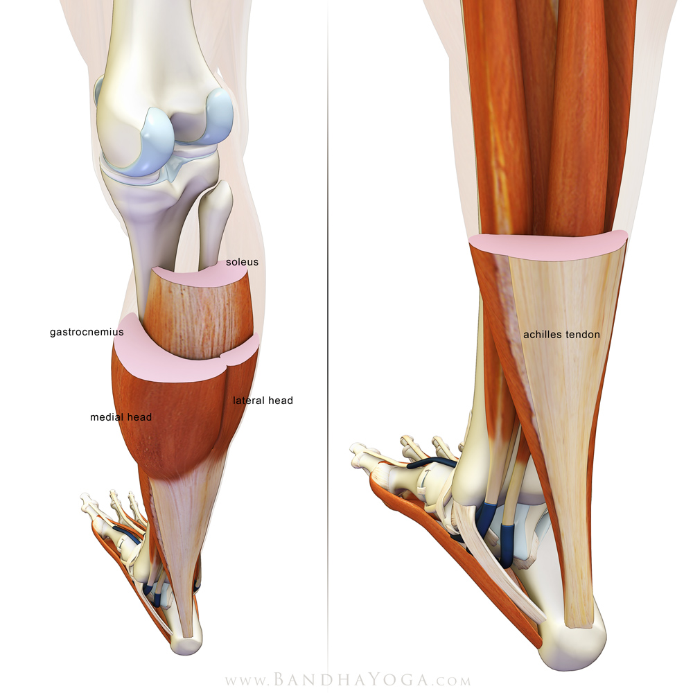
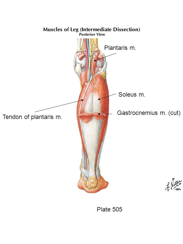
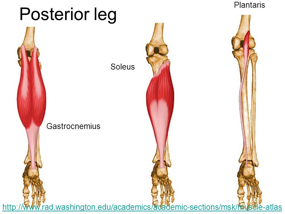
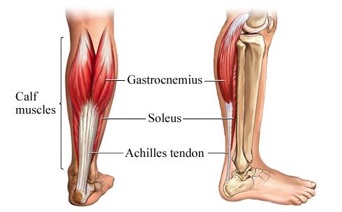
Posterior lower leg muscles: gastrocnemius, soleus, plantaris


Images:
Ankle bone: tibia, fibula, talus (sandwiched btw tibia & calcaneus), calcaneus (heel)

Lateral ligaments:


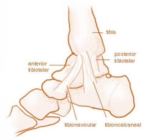
Medial ligament:

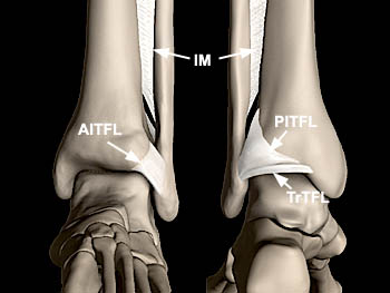
Tibiofibular syndesmosis:


 1 Anterior talofibular ligament; 2 anterior tibiofibular ligament;

3 anterior tibiofibular ligament
Lower leg muscles:


Video:

Sources:
Pictorial essay of ankle anatomy: http://www.ncbi.nlm.nih.gov/pmc/articles/PMC2855022/

Friday, March 18, 2016

superficial posterior compartment of leg

all insert @ the calcaneus thru achilles tendon: gastrocnemius, soles, plantaris


tibiofibular joint/syndesmosis


deltoid ligament

Prevents eversion
Ligament of the medial ankle.


Calcaneofibular ligament

One of the lateral ankle ligaments
Prevents inversion in neutral position

Location: "The calcaneo-fibular ligament is a ligament connecting the apex of the lateral malleolus to the posterolateral aspect of the calcaneus." (1) 

Significance: "Tenderness, swelling, and ecchymosis in the area inferior and posterior to the lateral malleolus is evidence of a calcaneo fibular ligament sprain." (1) 


---
Sources:
(1) http://www.orthopaedicsone.com/display/Main/Calcaneo+fibular+ligament+Surface+Anatomy

Posterior talofibular ligament

One of the ligaments of the lateral ankle
Strongest lateral ligament
Rarely injured


anterior talofibular ligament

One of the ligaments of the lateral ankle - most frequently injured w/ inversion injury.

Location: "The talofibular ligament is located between the anterior margin of the lateral malleolus (distal fibula) and the neck of the talus" (1)

Significance: "The talofibular ligament is the most common ankle ligament injured. It is often sprained during an acute ankle inversion. Inspect and palpate the ligament for tenderness, ecchymosis, and swelling to confirm a tear." (1) 

Anterior talo-fibular ligament

Talus head


Source:
(1) http://www.orthopaedicsone.com/display/Main/Anterior+talofibular+ligament+Surface+Anatomy