Monday, December 26, 2016

EKG height


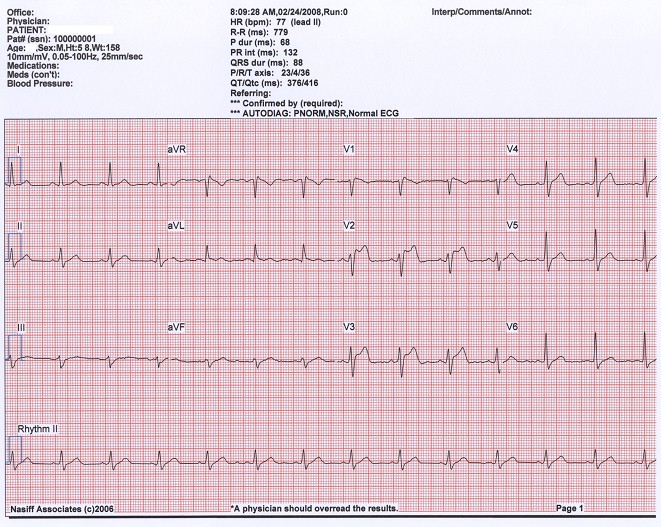
P waves in leads II and V1.
  •      If the P in lead II is greater than 2.5 mV (small boxes) right atrial enlargement (RAE) probably exists. 
  •       If the P wave in V1 is negative or biphasic, then LAE probably exists.  
    • This negative portion of the P wave in V1 should be more than 1 box wide and 1 box down to be considered significant.
PR segment
  • PR depression. The only classic significant PR segment abnormality that is encountered in the emergency department is PR depression.  This is most often seen in the setting of pericarditis.  Since there are multiple stages of pericarditis, these depressions are not always seen when this disease is present.
QRS
  • QRS width:
    • QRS >3ss (0.12 sec) = BBB 


  • QRS shape:
    • RBBB --- RSR' = rabbit ears = The “R” from Right and Rabbit = right BBB (RBBB) there should be an RSR’ in leads V1, V2 or V3.
    • LBBB --- a left BBB (LBBB) you need a deep, wide Q/S wave in these anterior leads.  This you will just have to memorize.


    •  QRS height:
      • LVH --- left ventricular hypertrophy (LVH)
        • A positive deflection in leads I or aVL greater than 11 mV. Mnemonics: 1 and L look like an 11 when they are side by side.
      

        • A value greater than 35 mV when you add the absolute values of the more negative of V1 or V2 plus the more positive of V5 or V6. Mnemonics: just have to look at the Q or S in leads V1 and V2 and see which is more negative.  Take the absolute size of that complex and add it to the larger R of V5 or V6.
        • Remember, only one criterion is sufficient to diagnose LVH.

      • ST segments:
        • you need to check systematically through all 12 leads of the EKG looking for ST elevations or depressions.  These findings are consistent with AMI or ischemia respectively.


      • T wave:
        • Specifically, you are looking for flipped T waves that are pointing in the negative direction.  This is also symbolic of coronary ischemia.  
       
        • Quickly glance at the shape of the T waves.  If they are sharp and pointy instead of nicely rounded, hyperkalemia may exist. 

          EKG width

          P wave width = 0.12 sec (3ss) (>0.12 or >3ss <=> atrial enlargement)
          PR interval (from the beginning of P wave to 1st deflection of QRS) = or <  0.20 sec (5ss = 1ls)  (if >0.20 or 5ss <=> 1st degree AV block)


          QRS width = 0.12 sec (3ss) (>0.12 or 3ss <=> bundle branch block (BBB)  (partial or full) OR the complex did not take the conduction pathway at all and had a ventricular origin as is seen in a premature ventricular complex or PVC)

          QTc width < 450sec => If >450msec, it may be a tip-off that electrolyte abnormalities exist or some toxin is present.  While this concept is more complex than depicted here, it is not so important and can be discussed later when looking at specific EKG’s


          ekg basics


          ekg axis

          LAD

          ekg rhythm

          P wave - precedes every QRS?
          P wave morphology - same shape? - originates in every SA node
          PR interval - same length?


          5 Study Techniques Every Clinical Student Should Know

          http://www.medscape.com/viewarticle/849951

          With the start of the new school year, 25,000 incoming medical students in the United States—and hundreds of thousands of students around the world—are wondering how best to study so they can succeed in classes, on board exams, and in the clinic. Fortunately, decades worth of neuroscience research has given us an entire toolkit of techniques, many of which you probably have not heard of before.
          Both of us have devoted much of our careers before and during medical school to assimilating and testing these cognitive techniques; the result of these efforts is our learning platform, Osmosis. Here we will highlight five of the most effective, neuroscience-backed study techniques that we've incorporated into Osmosis that every clinical student should know and how you can apply them.

          Technique 1: Test-Enhanced Learning

          Having taken dozens of high-stakes summative tests, ranging from class finals to the MCAT, SAT, and ACT, you've probably come to associate tests with the end of a learning experience rather than as part of it. Fortunately, over the past decade, researchers have been chipping away at this dogma; now, educators are beginning to view low-stakes formative tests as integral parts of the learning process. Testing has been shown to more effectively improve knowledge retention compared with less active forms of studying, such as rereading information or rewatching lectures. Thus, it is important to find opportunities to quiz yourself with flashcards and questions, ideally on a daily or weekly basis, to ensure that you're truly internalizing the material.
          In a New York Times commentary titled "How Tests Make Us Smarter," Professor Henry L. Roediger of the Washington University in St Louis further describes how active retrieval of information through testing strengthens the underlying knowledge. Equally important, however, is when you take these tests, which brings us to the next technique.

          Technique 2: Spaced Repetition

          First described in the 1880s by German psychologist Hermann Ebbinghaus, spaced repetition is hardly a novel technique. However, it's only now becoming widely adopted by students and teachers. The key concept is that spacing your studying and self-testing over time as opposed to massing, also known as "cramming," will flatten your forgetting curve and help you retain information longer.
          The reason cramming persists as a popular behavior is that it is often more effective in the short term. Pulling an all-nighter can certainly help you pass tomorrow's exam, but 1 month later, you will have forgotten much of that information. Given that learning medicine is more akin to an ultramarathon as opposed to a sprint, it would behoove you to space out your study sessions. There are many tools that can schedule these sessions for you, including Anki and Osmosis.
          It's important to note that spaced repetition does not just help with knowledge retention—it also helps with skill development, as this study on learning surgical procedures demonstrated.
          All this being said, recognizing potential issues with spaced repetition is also important. Given how dynamic medicine is (eg, guideline changes, pharmaceutical discoveries), you actually do not want to remember what you learn in school forever. That’s why you should use a spaced repetition system that updates you on these changes (something we’ve pioneered at Osmosis).

          Technique 3: Interleaved Practice

          Let's say that you want to learn concepts A, B, and C. The traditional model of education has you master each in turn through massed practice: AAABBBCCC. Interleaving mixes this up, for example: ABCBCABAC.
          Similar to spaced repetition, interleaving may not be as effective in the short term but is more effective in the long term. This is because massing often becomes a passive process, where your brain goes into cruise control and you default to mechanically applying knowledge; interleaving forces you to think through each concept every time and helps you figure out how they overlap and differ.
          Thus, for example, when you learn about the various diuretics, do not practice all the thiazides followed by all the loops; instead, interleave them. These first three techniques all fall under what UCLA psychology researcher Robert Bjork and his colleagues call "desirable difficulties." These are counterintuitive strategies that lead to reductions in short-term performance but improvements in long-term performance.

          Technique 4: Memory Associations

          Have you ever had difficulty recalling someone's name even though you remembered a seemingly obscure fact, such as where that person is from? This can in part be explained by the Baker-baker paradox, which essentially describes why it is easier to remember what someone does (baker) than what their name is (Baker). The name is simply a string of letters, whereas the profession is an evocative concept that helps you quickly form associations, such as your favorite baked good or the bakery near where you live. The more associations you can form to something you're trying to learn, the more likely you are to remember it in the future because there are more paths you can take to retrieve it.
          These associations can range from visually compelling images or "memory palaces," which is the basis for the education company Picmonic (which found up to a 331% improvement in memory 1 month after learning disease topics), to actual patient cases, which can be accessed through such tools as ReelDx Education and Celebrity Diagnosis. We recommend combining two or more of these tools to ensure that you're forming strong associations to the thousands of otherwise seemingly discrete facts that you encounter during your medical training.

          Technique 5: Fogg Behavior Model

          Now that you know a few techniques that are proven to help you learn more efficiently and remember longer, how do you actually make a habit of them? This is where the Fogg behavior model can transform your life (and help you counsel your future patients better). Stanford behavioral scientist BJ Fogg reduces behavior change to three variables: motivation, ability, and trigger. If you think about any behavior—exercising, quitting smoking, or studying using the techniques described above—you need a certain level of motivation and ability, followed by a trigger, to implement the behavior.
          For example, say you want to study new material and review past concepts on a daily basis. Your motivation is a desire to do well on class and board exams and, more important, when you see patients. You can increase your motivation through engaging such practices as gamification and social accountability (eg, study groups). You can increase your ability by using such tools as mobile apps that make this material readily accessible wherever you are. Finally, you can trigger the behavior through reminder emails, text messages, and push notifications.
          On this last point, the key is not to burn out on the triggers so that they do not have an effect in the future. One unique way to decrease the chance of this happening is by making each trigger relevant by syncing it to your individual curriculum and schedule—so you get, for example, practice questions on gram-positive bacteria when you're actually learning about them as opposed to randomly. We've previously written in the Annals of Internal Medicine about how we've implemented this using an intelligent recommendation systemic, similar to what Amazon and Netflix use.

          Further Reading

          If you're interested in reading more about the techniques above, we suggest you take a look at three books in particular: Make It Stick: The Science of Successful Learning, Moonwalking With Einstein: The Art and Science of Remembering Everything, and Learning Medicine: An Evidence-Based Guide.

          Bringing It All Together

          Now that you know about these effective strategies, how do you make sure you actually apply them? Fortunately, the behavior change model above can help you implement these and other behaviors in your life. We also recommend checking out the many educational tools that enable you to use one or more of these techniques, so that you are more efficient with your limited time.
          Good luck!
          Normal EKG


          ekg rate

          Mnemonics 300-150-100-75-60-50
          300/n


          300/1 = 300
          300/2 = 150
          300/3 = 100
          300/4 = 75
          300/5 = 60
          300/6 = 50


          Tuesday, December 20, 2016

          Resources list


          Resources List Compiled by Qt

          EKG:
          EKG Basic steps: https://kchemekg.wordpress.com/ekg-test-home/
          EKG Basic steps: http://rebelem.com/ecg-basics/
          EKG Cases: http://hqmeded-ecg.blogspot.com/

          Clinical guidelines resources:
          Medscape www.medscape.com (my personal favorite resource, FREE but you have to register, highly organized and pretty updated).

          Uptodate www.uptodate.com (very expensive $$$, lots of entries, some even said that they contain more articles than medscape)

          Dynamed

          Essentialevidenceplus www.essentialevidenceplus.com (not really my favorite because it looks like it's a big database of original or cochrane reviews but it's not organized for quick review like uptodate or medscape. Yet, it has lots of entries. You can try it out for free in 30 days to get a feel of it.)


          Gastroenterology (GI):
          American College Gastroenterology - Clinical Guidelines: http://gi.org/clinical-guidelines/clinical-guidelines-sortable-list/ (very useful resource for several key GI conditions)

          GI Pathology association - GI pathology CME courses: http://usgips.com/resources/gi-pathology-courses/ (probably too high a level for general non-pathology GI NP students like me)


          Suture skill:
          http://www.lectoro.com/index.php?action=search&ytq=Duke%20Suture%20Skills%20Course
          http://www.theapprenticedoctor.com/elearning/suture/


          Learning skills:
          Online learning: https://onlinelearninginsights.wordpress.com/2012/09/28/five-step-strategy-for-student-success-with-online-learning/ 
          Study tips: https://meded.ucsd.edu/index.cfm/ugme/oess/study_skills_and_exam_strategies/how_to_study_actively/

          Surgery:
          Ambulatory: http://fitsweb.uchc.edu/student/selectives/Luzietti/Ambulatory_Surgery_mainpage.htm
          Old texts: http://www.meb.uni-bonn.de/dtc/primsurg/index.html
          Pt edu for surgery: https://www.facs.org/education/patient-education/patient-resources/operations


          Case study:
          http://reeldx.com/
          http://www.celebritydiagnosis.com/ 


          Endocrine:
          https://medicine.yale.edu/intmed/endocrin/patient/Yale%20National%20F_50135_1095_4813.pdf

          Opthalmology:
          http://webeye.ophth.uiowa.edu/eyeforum/cases/case28.htm

          Pt edu:
          http://patient.info/


          Sunday, December 11, 2016

          learning resources

          Online learning resources:

          https://onlinelearninginsights.wordpress.com/2012/09/28/five-step-strategy-for-student-success-with-online-learning/

          Monday, December 5, 2016

          neck strain xray = straightening of cervical curvature

          What Is Straightening of the Normal Cervical Lordosis? Can It Cause Problems?

          what is straightening of the normal cervical lordosis
          The fact that the neck has a normal curve is an important and often overlooked issue. In many patients with neck pain, this curve goes away, leading to biomechanical problems that can cause degenerated discs. Now a new study shows that it can also cause decreased blood flow to the back of the brain!

          What Is Straightening of the Normal Cervical Lordosis?

          If you read a handful of neck X-ray reports, you’ll likely see that the radiologist has said on a few that there is “straightening of the normal cervical lordosis.” To understand what this is, you have to understand how the spine is constructed.what is straightening of the cervical lordosis
          When the normal curve is lost, there are a number of terms used by the radiologist:
          • Straightening of the normal cervical lordosis
          • Loss of cervical lordosis
          • Straightening of the cervical lordosis
          Your neck, upper back, and low back all have counterbalanced curves. One goes one way, and the adjoining curve goes the opposite way. When the curve points toward the front, it’s called a lordosis and toward the back, it’s called a kyphosis. These curves balance the force of the head and allow you to stand with minimal muscle energy. They also equally distribute the forces between the front of the individual vertebrae, where the disc is located, and the back of the verterbrae, where the facet joints live.

          Why Is Straightening of the Normal Cervical Lordosis a Big Problem?

          loss of cervical lordosisWhen you lose the normal neck curve, the weight of the head is no longer balanced by the opposing curve (kyphosis) in the upper back. This causes muscle power to be needed to keep the head upright. For example, in the picture to the right, the head is being held up by the muscles at the back of the neck, leading to overload where those muscles attach to the skull, causing headache pain. This also causes more weight to be transferred to the discs between the vertebrae, which can lead to more wear-and-tear disc degeneration.

          How Does Straightening of the Normal Cervical Lordosis Happen?

          One of the more common causes of straightening of the normal cervical lordosis is car crashes where the neck ligaments are damaged. Other causes are more insidious, like looking down all the time to interact with a cell phone, tightness in the chest wall and psoas from sitting too much, or just getting older.

          New Research on Straightening of the Normal Cervical Lordosis

          While we’ve known that straightening of the neck curve was bad news, a new study just upped the ante of bad stuff that happens when you have this problem. This research looked at 30 patients with and 30 matched controls without loss of cervical lordosis. Doppler ultrasound was used to measure the blood flow through the vertebral artery. This important blood vessel courses through holes in the neck bones, so it could be vulnerable if the relationships between the neck bones change. In the patients with loss of the normal neck curve, the diameter of the artery was small with less and slowed blood flow through the vessel. Yikes! Given that this is one of the major sources of blood flow to the brain, this could be a huge future problem for teenagers who have been glued to their phone since childhood!

          How Can You Treat Straightening of the Normal Cervical Lordosis?

          From a conservative-therapy standpoint, there are some nice programs specifically designed to get the curve back. One is used by chiropractors and uses special traction machines to fix the curve. Physical therapists can also work on stretching out and releasing the tight muscles in the chest and pelvis. If the neck ligaments are damaged, then highly precise injections into these ligaments using platelets or stem cells may help. Surgery is usually not recommended as a viable solution.
          The upshot? We’ve known that the neck curve is a big deal in helping avoid structural overload and degeneration of muscles, tendons, and discs, but this new study is a bit scary. The fact that the blood supply to the brain is also impacted by this change is a big 


          http://www.regenexx.com/what-is-straightening-of-the-normal-cervical-lordosis/

          tibia vs fibula


          o remember how to distinguish the fibula from the tibia, consider the “fib” and “l” parts of the word fibula. To tell a small lie is to fib. The fibula is smaller than the tibia, and is also lateral to it.
          To remember where the talus is located, think that the talus is on top of the foot and articulates with the tibia.
          http://www.judinath.com/tipstricksforana.html

          Fibula vs. tibia: which is the bigger one

          People talk of telling just a “little white lie”, or a “little fib”:
          Therefore, the fibula is smaller.
          • Alternatively: TIBia is the Thicker Inner Bone.
          • Alternatively: “Little lat FIB, big fat TIB”.

          Tibia & Fibula

          Fragile: The Fibula is thin & fragile
          Thick: Tibia is thick 

          buckle fx

          Torus fractures, also known as buckle fractures, are incomplete fractures of the shaft of a long bone that is characterised by bulging of the cortex. They result from trabecular compression from an axial loading force along the long axis of the bone. They are usually seen in children, frequently involving the distal radial metaphysis.

          Epidemiology

          These type of fractures are more common in children, especially aged 5-10 years, due to the elasticity of their bones.

          Pathology

          Cortical buckle fractures occur when there is axial loading of a long bone. This most commonly occurs at the distal radius or tibia following a fall on an outstretched arm; the force is transmitted from carpus to the distal radius and the point of least resistance fractures, usually the dorsal cortex of the distal radius.

          Radiographic features

          Plain radiograph
          • distinct fracture lines are not seen
          • subtle deformity or buckle of the cortex may be evident
          • in some cases, angulation is the only diagnostic clue

          Treatment and prognosis

          They are self-limiting and typically do not require operative intervention, although a manipulation may be required if the angulation is severe. Sometimes a cast may be applied, but often a splint is all that is required with a period of rest and immobilisation.

          History and etymology

          The term torus is derived from the Latin word tori which means protuberanceA torus is the convex portion of the upper part of the base of a Greek column (figure 1), and resembles the appearance of the cortical buckling seen in the "column" of bone which has been fractured in the pattern discussed in this article.

          radiology

          https://radiopaedia.org/
          http://www.radiologymasterclass.co.uk/tutorials/musculoskeletal/x-ray_trauma_upper_limb/elbow_fracture_x-ray

          bursa

          source: http://study.com/academy/lesson/bursa-definition-anatomy.html

          Bursae are fluid-filled sacs located around a joint that cushion bones when muscles, tendons, or skin rub against the bone. They are very important in movement of the bones because they help reduce friction. Without them, movement would be difficult and even painful. Now let's try and relate this to something you may know about.
          Let's say your friend told you about an incident that happened last week in the factory where he works. One of the machines made a very loud grinding sound and then suddenly stopped working. On inspection, your friend tells you that the washers that lie between the metal parts of one of the machines broke. The grinding was the sound made when metal rubbed on metal. In this example, the washers were crucial to having the machine work. Likewise, the bursae are crucial for having the joint work properly

          Anatomy

          Wherever there are tendons moving across a bony surface, there is a bursa. There are approximately 150 bursa found in the body. Most are present at birth, but a bursa may form in an area where there is friction. Bursae are filled with a thin layer of thick fluid (like the consistency of a raw egg white) and a lining of synovial fluid, which also works as a 'shock absorber' for the joint and supplies molecules to the cartilage. This is known as 'dissipating force'. Normally, the bursae are thin and provide a very slippery surface so there will be very little friction when there is movement of the joint.
          Think of a slide on a playground. If a child has a piece of wax paper and sits on it to slide, they go down very quickly don't they? This would represent the function of a bursa. But if the child has on shorts and tries to slide down - the skin can cause more friction and the child can hardly move down the slide. If no bursae are present at the joint, bending your elbow would be like trying to go down a slide with shorts on.

          Sunday, December 4, 2016

          pathology images

          http://peir.path.uab.edu/library/

          nephrolithiasis

          diet: http://kidneystones.uchicago.edu/how-to-eat-a-low-oxalate-diet/

          My notes for Microcytic anemia

          Microcytic => IRON studies. Look at IRON => Then Look at TIBC&Ferritin
          1.       Low ironmicrocytic anemia =
          a.       Iron deficiency:  
                                                                         i.      Labs:
          1.       Low Serum Fe
          2.       High TIBC
          3.       Low Ferritin levels are generally accepted as reliable single indicators of the presence of iron deficiency.
                                                                       ii.      Interpretation: The liver produces a lot of TIBC trying to maximize little Fe that is available. Not enuf Fe to store in Ferritin (Low ferritin). The body tries to produce lots of TIBC/transferritin to absorb all little Fe there is to absorb.
                                                                     iii.      Dx: Low iron deficiency
                                                                     iv.      Tx: here’s the iron pill, have a nice day!
          b.       Chronic disease anemia:
                                                                         i.      Labs:
          1.       Low serum Fe
          2.       Low TIBC
          3.       High Ferritin
                                                                       ii.      Interpretation: The body holds Fe intracellularly via Ferritin to keep Fe away from offending tumor/bacteria. The body produces more Ferritin in order to store and keep all Fe there is that is required for the synthesis of offending organism or tumor as they use Fe for their pathosynthetic disease.
                                                                     iii.      Dx: chronic disease anemia
                                                                     iv.      Tx: address underlying causes.
          2.       Normal iron microcytic anemia
          a.       Labs:
                                                                         i.      Normal Fe
                                                                       ii.      High TIBC
          b.       Interpretation:
                                                                         i.       Normal Serum Fe => there’s sth happening that the body is n

          c.       Dx: do Hgb electrophoresis to r/o thalassemia (genetically abnormal Hgb), do BMP, pregnancy or use of hormonal contraception

          ~Qt

          Friday, December 2, 2016

          drug stem books

          http://www.who.int/medicines/services/inn/StemBook_2011_Final.pdf

          Thursday, December 1, 2016

          Top 4 Common Extensor Surface Skin Conditions

          Source: https://www.verywell.com/extensor-surface-1069321
          An extensor surface is the skin on the opposite side of a joint. In other words, it's the surface of an extensor muscle: a muscle that flexes when it bends and extends when it straightens.
          Take the elbow, for example. When you bend your elbow, the skin on the inner forearm and inner upper forearm touch. That's known as a flexor surfaceor the parts of the skin that touch when a joint bends. The flexor surface of the leg is the back of the leg.
          The extensor surface of the arm is the outer arm, or the parts of the skin on the opposite side of the joint. Extensor surfaces do not touch. The legs are another example. The extensor surface of the leg is the front of the leg. The flexor surface is the back of the leg.

          Types of Extensor Surfaces

          Extensor muscles exist in the arms, hands, legs and feet. They specifically include:
          • Arm and shoulder
          • Forearm and elbow
          • Hand and wrist
          • Fingers
          • Thigh and hip
          • Leg and knee
          • Toes

          Extensor Surfaces & Skin Conditions

          There are several different skin conditions that seem to favor taking hold of extensor surfaces. These conditions include psoriasis, erythema multiforme, eczema and dermatitis herpetiformis.

          Psoriasis

          Psoriasis, one of the most common skin diseases, involves the overproduction of keratinocytes in the epidermis, which, in turn, increases the cell turnover rate. There are several different types of psoriasis, all of which can appear nearly anywhere on the body.
          Chronic stationary psoriasis, also referred to as psoriasis vulgaris, is the most common type of psoriasis. It commonly appears on extensor surfaces such as the knees and elbows. Plaque psoriasis almost exclusively affects extensor surfaces, like the elbows.
          Psoriatic arthritis is a type of arthritis associated with psoriasis.
          Symptoms of each condition develop separately, and in most cases psoriasis symptoms precede arthritis symptoms. Any joint on the body can be affected by psoriatic arthritis. Symptoms of psoriatic arthritis include stiffness of joints, fatigue, swelling of the fingers and toes, tendinitis, lower back pain and even conjunctivitis.

          Erythema Multiforme

          The exact cause of the skin condition erythema multiforme is unknown, but it's believed to be caused by an allergic reaction or infection. Certain medications including barbiturates, penicillin, phenytoin and sulfonamides can trigger a reaction, as can illnesses like herpes simplex and mycoplasma.
          There are many symptoms associated with erythema multiforme, many of which are related to the skin. Specifically, round, bullseye-shaped lesions appear on extensor surfaces like the arms and legs.

          Eczema

          Eczema is a type of skin condition characterized by patches of rough, inflamed skin that seems to overwhelmingly favor flexor surfaces, but it can appear on extensor surfaces.
          In cases of nummular eczema, circular, well-defined, scaly plaques appear on extensor surfaces including the arms, legs and hips.

          Dermatitis Herpetiformis

          Dermatitis herpetiformis is a somewhat uncommon type of extremely itchy, persistent rash. It's thought to appear due to an autoimmune disorder, and recent research suggests a link between the skin condition and celiac disease. It's characterized by itchy bumps and blisters that appear on extensor surfaces such as the elbows and knees.

          Wednesday, November 30, 2016

          Jail medicine

          http://www.jailmedicine.com/

          Cellulitis vs. Abscess

          Cellulitis is a diffuse infection of the soft tissues with no localized area of pus amenable to drainage. The affected area is described as indurated (i.e., warm, red, and swollen). It is also painful. A component of lymphangitis (infection involving the lymphatics) is indicated by red streaking, progressing proximally from the affected area.

          An abscess is a localized collection of pus, often with a component of surrounding cellulitis (with the above signs). One sign of an abscess is an area of fluctuance; that is, when you apply gentle digital pressure over the area, you can push and feel a “give,” indicating the presence of fluid underneath. Another sign is that an abscess often seems to “point;” that is, the skin starts to thin from the pressure of the fluid underneath. The distinction between cellullitis and abscess is important. The main treatment for an abscess is incision and drainage (cutting into the abscess and widely opening the abscess cavity). Cellulitis does not warrant this intervention.

          surgeries for non-surgeons

          http://practicalplasticsurgery.org/

          GAL


          skin infxn

          http://www.forbes.com/sites/judystone/2015/03/18/choosing-an-antibiotic-for-skin-infections-whats-best/#197415c17065

          antibiotics

          Source: http://www.microbiologynutsandbolts.co.uk/the-bug-blog/-how-to-remember-antibiotic-spectrums-of-activity
          I am often asked “how can doctors remember the different
          antibiotic spectrums of activity”? This is a difficult task as there are so many. Personally I have never found lists of medical information that helpful, I would much rather come up with a mnemonic. However, I’m stumped for a mnemonic to remember antibiotic spectrum of activity, so a list it must be. What I have done though is reduce a very long list into six short related lists, which I hope are easier to remember.

          NOT knowing the spectrum of activity of the antibiotic they are prescribing is high up there on the list of situations when doctors make a mistake. NOTknowing leads to patients being given antibiotics which have no activity against the bacteria which are known or suspected to be the cause of an infection. My current pet hate is the use of Teicoplanin to treat UTIs (see Teicoplanin blog...) but I also come across patients being treated for Pseudomonas spp. with Co-amoxiclav which has no activity against Pseudomonas spp., or an anaerobic infection being treated with both Co-amoxiclav AND Metronidazole when either alone would be enough. Sometimes it seems doctors are just guessing or picking their favourites!

          Remembering the 6 lists below will help to ensure patients are on an antibiotic that is active against their infection.
          1. Active against Gram-positive and Gram-negative bacteria (Broad-spectrum antibiotics)
          2. Active against anaerobes
          3. Active against Pseudomonas spp.
          4. Active ONLY against Gram-positive bacteria
          5. Active ONLY against Gram-negative bacteria
          6. Active against “non-culturable” bacteria

          Lists 1 and 2
          Antibiotic Spectrum of Activity Lists 1&2
          Click for larger image
          Example A: A patient has a perforated appendix. Which antibiotics could be used to cover the mixed bowel flora that will have leaked into the patient’s peritoneum? Any of Co-amoxiclav, Piptazobactam, Ertapenem, Imipenem or Meropenem could be used alone as they are broad spectrum AND cover anaerobes. If Cefuroxime, Ceftriaxone or Cefotaxime were used then Metronidazole would need to be added to cover the anaerobes. You would not use Cefuroxime, Ceftriaxone or Cefotaxime with Clindamycin as this poses too high a risk for Clostridium difficile associated diarrhoea (CDAD).

          Example B: In patients with serious penicillin allergies Beta-lactams including Co-amoxiclav, Piptazobactam, Ertapenem, Imipenem or Meropenem are contraindicated. Combinations of antibiotics are therefore needed to cover the gut flora, such as Teicoplanin OR Vancomycin (which only have Gram-positive activity) PLUS Metronidazole (which only covers anaerobes) PLUS either Ciprofloxacin ORGentamicin (both of which have excellent Gran-negative activity).

          List 3
          Antibiotic Spectrum of Activity List 3
          Click for larger image
          Example C: A Cystic Fibrosis (CF) patient has an exacerbation of their CF caused by Pseudomonas aeruginosa. What antibiotics would they be treated with? They would be treated with a combination of antibiotics active against Pseudomonas aeruginosasuch as IV Meropenem PLUS IV Tobramycin PLUSnebulised Colistin.
          Why a combination of antimicrobials? Combination antimicrobials are used to try and prevent further development of resistance (Pseudomonas aeruginosais notorious for acquiring new resistance mechanisms), in non-CF patients two antimicrobials are usually enough. It is unlikely that bacteria will become resistant to two or more antimicrobials at the same time therefore resistance is less likely to occur. Even if the bacteria do become resistant to
          one of the antimicrobials the other agent(s) will be able to kill the bacteria.

          List 4 and 5
          Antibiotic Spectrum of Activity Lists 4&5
          Click for larger image
          Example D: A patient has a skin and soft tissue infection but also has a severe allergy to penicillins. Which antibiotics could be used to treat them? Skin and soft tissue infections are almost always caused by Gram-positive bacteria such as Staphylococcus aureus or Beta-haemolytic streptococci therefore Vancomycin, Teicoplanin, Linezolid and Daptomycin would all be capable of treating the infection. It may sound obvious but you cannot use an antimicrobial only active against Gram-negatives to treat a Gram-positive, therefore Ceftazidime, Colistin or Aztreonam would definitely not be appropriate as they do not have any activity against the Gram-positive causes. Broad-spectrum antibiotics would treat the infection but it would be unnecessary as the bacteria are known to be Gram-positive. Broad spectrum antibiotics in this case might lead to unwanted side effects such as CDAD.

          List 6
          Antibiotic Spectrum of Activity List 6
          Click for larger image
          Example E: A patient has pneumonia caused by Mycoplasma
          pneumoniae
           (a non-culturable bacterium). What antibiotic should they be treated with? Erythromycin, Clarithromycin, Azithromycin, Ciprofloxacin, Levofloxacin and Doxycycline all could be used to treat this patient.

          If you can learn these six lists it starts to become much easier to understand why certain antibiotics are chosen to treat certain infections. It also enables you to choose alternatives when patients are allergic to first line antibiotics found in guidelines. Downloada handy postcard of the 6 lists and keep it in your book Microbiology Nuts and Bolts.

          Give it a try and see if it helps. Pass it on too. If you can think of any easy way of remembering them then let me know, I’m always keen to know how others have learned their microbiology.